Research focus
In our research projects, we aim at utilizing microtechnology and applied mathematics to create new forums that can transfer the unique physical/chemical phenomenon into platforms for biomedical applications such as tissue engineering, disease diagnostics or therapeutics.
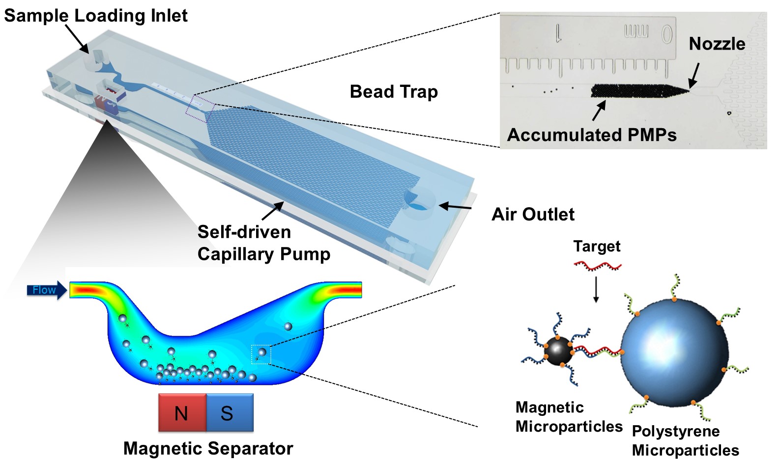 Microfluidic
Bead
Trap is
a dipstick-type bar visible by naked eyes
for
simple and quantitative detection of oligonucleotides.
In recent years, visual detection, as a simple, direct and
rapid method, has been developed in many platforms. However,
while convenient, current platforms for visual detections
are mostly limited to qualitative results (yes or no
signal). For quantitative measurement, the visual signal
needs to be converted into optical/fluorescence intensity
measured by lateral flow strip reader, or spectral
absorbance read by UV-Vis spectrometer. As a result,
cumbersome, bulky instruments and power supply are
inevitable, which creates technical hurdles for detection
and analysis in resource-limited settings. Thus, we
use magnetic
microparticles (MMPs) and polystyrene microparticles (PMPs)
that are
connected and form
MMPs-targets-PMPs when target
oligonucleotides are present,
leaving free PMPs
with a number
inversely proportional to the
amount of targets.
Using a capillary flow-driven
microfluidic circuitry consisting of a
magnetic
separator to remove the
MMPs-targets-PMPs,
the free
PMPs can be
trapped at the narrowing nozzle in
downstream, forming a visual bar
quantifiable
based on the
length of PMP
accumulation (Zhao, Lab
Chip, 2017).
We anticipate that this method can provide a simple,
sensitive approach achieving visual detection and direct
quantification of lead contamination on a power-free and
instrumental-free platform.
Microfluidic
Bead
Trap is
a dipstick-type bar visible by naked eyes
for
simple and quantitative detection of oligonucleotides.
In recent years, visual detection, as a simple, direct and
rapid method, has been developed in many platforms. However,
while convenient, current platforms for visual detections
are mostly limited to qualitative results (yes or no
signal). For quantitative measurement, the visual signal
needs to be converted into optical/fluorescence intensity
measured by lateral flow strip reader, or spectral
absorbance read by UV-Vis spectrometer. As a result,
cumbersome, bulky instruments and power supply are
inevitable, which creates technical hurdles for detection
and analysis in resource-limited settings. Thus, we
use magnetic
microparticles (MMPs) and polystyrene microparticles (PMPs)
that are
connected and form
MMPs-targets-PMPs when target
oligonucleotides are present,
leaving free PMPs
with a number
inversely proportional to the
amount of targets.
Using a capillary flow-driven
microfluidic circuitry consisting of a
magnetic
separator to remove the
MMPs-targets-PMPs,
the free
PMPs can be
trapped at the narrowing nozzle in
downstream, forming a visual bar
quantifiable
based on the
length of PMP
accumulation (Zhao, Lab
Chip, 2017).
We anticipate that this method can provide a simple,
sensitive approach achieving visual detection and direct
quantification of lead contamination on a power-free and
instrumental-free platform.
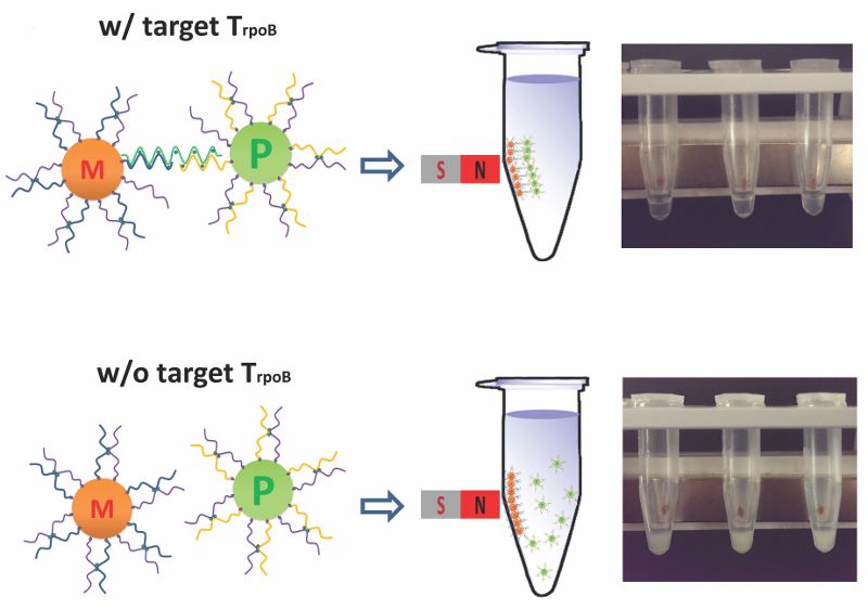 Bead
based biosensor is
to provide a rapid and simple approach for detection of
biomolecules such as nucleic acid or protein biomarkers.
Recently, many biomarkers present in the blood stream were
recently found to show promise for cancer classification
and prognostication. However, the complex environment of
blood has created significant challenge for on-site
detection. Using functionalized microparticles, we explore
the possiblity of visual detection of nucleic acids. With
the use of magnetic microparticles and polystyrene
microparticles, the visual readouts have been resorted to
Mie scattering (Zhao, Analyst, 2015), which provide greatly
enhanced extinction coefficient based on the magnetic
extraction and the stability of mono-dispersion, enabling
sensitive, multiplexed assays and ability for handling
complex fluids, such as whole blood, in a single assay.
Thus, by satisfying many of the requirements of
point-of-care detection, we envision that this method will
be applicable to healthcare and environmental monitoring in
resource-limited settings in the future.
Bead
based biosensor is
to provide a rapid and simple approach for detection of
biomolecules such as nucleic acid or protein biomarkers.
Recently, many biomarkers present in the blood stream were
recently found to show promise for cancer classification
and prognostication. However, the complex environment of
blood has created significant challenge for on-site
detection. Using functionalized microparticles, we explore
the possiblity of visual detection of nucleic acids. With
the use of magnetic microparticles and polystyrene
microparticles, the visual readouts have been resorted to
Mie scattering (Zhao, Analyst, 2015), which provide greatly
enhanced extinction coefficient based on the magnetic
extraction and the stability of mono-dispersion, enabling
sensitive, multiplexed assays and ability for handling
complex fluids, such as whole blood, in a single assay.
Thus, by satisfying many of the requirements of
point-of-care detection, we envision that this method will
be applicable to healthcare and environmental monitoring in
resource-limited settings in the future.
 Microtechology is
an essential tool allowing spatial modulations such as
physical stimuli or diffusion gradient of chemicals to
control the biological systems. With
the enhanced efficiency and efficacy, it has been used to
optimize the design principle in electrochemical sensor
(Garcia, Chen, JALA, 2009). The dominance of surface tension
in microscale can also be exploited in the droplet based
fluidic systems (Chen, et al, J. Micromech. Microeng.,
2007), or a separation mechanism for biological entities
(Wong, Chen, et al, Anal. Chem., 2011). Integrating with
cell biology, the interface between cell-adhesive and
non-adhesive substrate also play as an powerful stimulus to
direct this intrinsic motility of cells (Chen, et.al, Circ
Res, 2012; Chen, et.al, Biomaterials, 2012). Overall, it
creates a new venue with great flexibility and versatile
application for a variety of experimental scenarios.
Microtechology is
an essential tool allowing spatial modulations such as
physical stimuli or diffusion gradient of chemicals to
control the biological systems. With
the enhanced efficiency and efficacy, it has been used to
optimize the design principle in electrochemical sensor
(Garcia, Chen, JALA, 2009). The dominance of surface tension
in microscale can also be exploited in the droplet based
fluidic systems (Chen, et al, J. Micromech. Microeng.,
2007), or a separation mechanism for biological entities
(Wong, Chen, et al, Anal. Chem., 2011). Integrating with
cell biology, the interface between cell-adhesive and
non-adhesive substrate also play as an powerful stimulus to
direct this intrinsic motility of cells (Chen, et.al, Circ
Res, 2012; Chen, et.al, Biomaterials, 2012). Overall, it
creates a new venue with great flexibility and versatile
application for a variety of experimental scenarios.
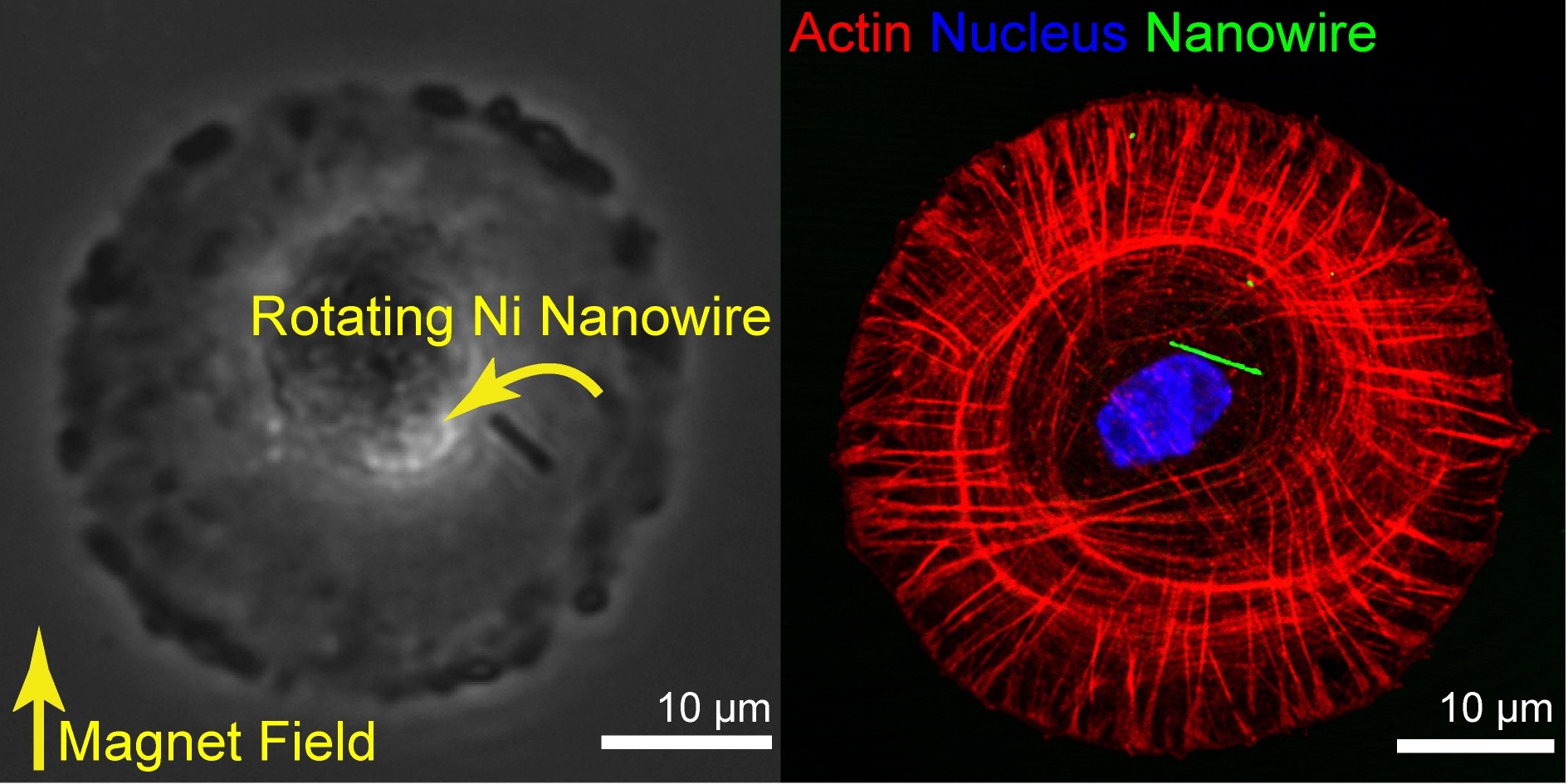
 Cell
mechanics plays
a central role in regulating cell proliferation,
differentiation, and morphogenesis.a central role in regulating cell proliferation,
differentiation, and morphogenesis.
How does cell mechanics regulate
cellular behavior? To probe this question, we cultured cells
on microengineered substrate which defines alternating
stripes of cell adhesive and non-adhesive substrate. We
found that cellular mechanical stress accumulated at the
substrate interfaces triggers an inherent left-right
asymmetry of cells. Based on it, cells preferentially
turn right on
migration across the interfaces, eventually leading to
multicellular structures with left-right asymmetry (Chen,
et.al, Circ Res, 2012). Importantly, this finding is the
first demonstration that tissue morphology with left-right
asymmetry can be formed from single-cell organizer which has
left-right preference. Also, although cells have been cultured
for decades, left-right biased migration in cultured adult
cells is rarely seen. Given that mechanical inductions are
generally absent in the conventional cultures, our findings
suggest that it is because cytoskeletal remodeling like
those in our experiments was not activated in the
traditional way.
Cell
mechanics plays
a central role in regulating cell proliferation,
differentiation, and morphogenesis.a central role in regulating cell proliferation,
differentiation, and morphogenesis.
How does cell mechanics regulate
cellular behavior? To probe this question, we cultured cells
on microengineered substrate which defines alternating
stripes of cell adhesive and non-adhesive substrate. We
found that cellular mechanical stress accumulated at the
substrate interfaces triggers an inherent left-right
asymmetry of cells. Based on it, cells preferentially
turn right on
migration across the interfaces, eventually leading to
multicellular structures with left-right asymmetry (Chen,
et.al, Circ Res, 2012). Importantly, this finding is the
first demonstration that tissue morphology with left-right
asymmetry can be formed from single-cell organizer which has
left-right preference. Also, although cells have been cultured
for decades, left-right biased migration in cultured adult
cells is rarely seen. Given that mechanical inductions are
generally absent in the conventional cultures, our findings
suggest that it is because cytoskeletal remodeling like
those in our experiments was not activated in the
traditional way.
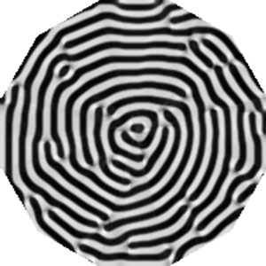
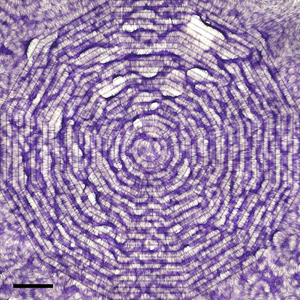 Tissue
formation is
the central core in many biological organizations, with
implications for many diseases such as spinal cord injury or
heart diseases.
However, how can we guide the cells to rebuild the damage or
missing tissue? When
rebuilding a tissue replacement,
self-organization also creates inherent challenge that may
frustrate and disorganize the artificial attempts. To work
in concert with self-organization rather than against it, we
use mathematical models that formulate the Turing's
reaction-diffusion mechanism as observed in many
developmental process. By modeling the kinetics of a pair of
growth factor, slowly-diffusing activator and its
rapidly-diffusing inhibitor, together with cellular
activity, we can reproduce the nonlinear dynamics that
results in specific pattern formations representing the
tissue development. Importantly, small difference in the
initial cell distribution (Chen, et.al, Biomaterials, 2012)
and cellular motility (Chen, et.al, Interface Focus, 2012)
can be integrated and amplified into changes in global
tissue morphology, shows a completely different route for
designing and reconstructing tissue with minimum engineering
efforts.
Tissue
formation is
the central core in many biological organizations, with
implications for many diseases such as spinal cord injury or
heart diseases.
However, how can we guide the cells to rebuild the damage or
missing tissue? When
rebuilding a tissue replacement,
self-organization also creates inherent challenge that may
frustrate and disorganize the artificial attempts. To work
in concert with self-organization rather than against it, we
use mathematical models that formulate the Turing's
reaction-diffusion mechanism as observed in many
developmental process. By modeling the kinetics of a pair of
growth factor, slowly-diffusing activator and its
rapidly-diffusing inhibitor, together with cellular
activity, we can reproduce the nonlinear dynamics that
results in specific pattern formations representing the
tissue development. Importantly, small difference in the
initial cell distribution (Chen, et.al, Biomaterials, 2012)
and cellular motility (Chen, et.al, Interface Focus, 2012)
can be integrated and amplified into changes in global
tissue morphology, shows a completely different route for
designing and reconstructing tissue with minimum engineering
efforts.